
The Definition and Importance of the Compound Microscope
A compound microscope is an optical instrument designed to magnify small objects by using multiple lenses to achieve high-resolution imaging. Unlike simple microscopes, which use a single lens, the compound microscope employs a system of objective and eyepiece lenses to provide significantly greater magnification and clarity. This tool is indispensable in scientific research, medical diagnostics, and educational settings, enabling users to observe details invisible to the naked eye. The definition of compound microscope highlights its dual-lens system, which allows for magnification levels ranging from 40x to 1000x, making it ideal for studying cellular structures, microorganisms, and intricate biological specimens.
Why It Matters:
- Scientific Discovery: Revolutionized biology, microbiology, and materials science.
- Medical Diagnostics: Essential for analyzing blood samples, tissue sections, and pathogens.
- Educational Tool: Bridges theoretical knowledge with hands-on experimentation in schools and universities.
Pro Tip: Ensure your compound microscope is calibrated correctly to avoid distorted images.
A Brief History of the Compound Microscope
The compound microscope traces its origins to the late 16th century, when Dutch spectacle makers Zacharias Janssen and his father Hans are credited with creating the first prototype. By the 17th century, scientists like Antonie van Leeuwenhoek and Robert Hooke refined its design, leading to breakthroughs in microbiology. The 19th century saw significant advancements in lens quality and mechanical stability, culminating in the modern compound microscope we use today.
Key Milestones:
- 1595: Janssen’s invention of the two-lens system.
- 1665: Hooke’s Micrographia documented observations using a compound microscope.
- 1830s: Improved achromatic lenses reduced chromatic aberration.
How It Evolved:
Modern compound microscopes now integrate digital imaging, automated focusing, and LED lighting for enhanced usability.
Key Components of a Compound Microscope
Understanding the parts of a compound microscope is crucial for optimal use. Each component plays a specific role in achieving clear, magnified images.
Main Parts:
- Objective Lenses: Provide primary magnification (4x, 10x, 40x, 100x).
- Eyepiece (Ocular Lens): Magnifies the image produced by the objective (usually 10x).
- Stage: Holds the specimen slide in place.
- Condenser: Focuses light onto the specimen for brighter, sharper images.
- Illuminator: LED or halogen light source for specimen illumination.
- Coarse/Fine Adjustment Knobs: Adjust focus for clarity.
- Diaphragm: Controls light intensity and contrast.
Pro Tip: Clean lenses with specialized microfiber cloths to prevent scratches and smudges.
How to Use a Compound Microscope Effectively
Operating a compound microscope requires careful technique to avoid damaging the instrument or specimen. Follow this step-by-step guide:
- Prepare the Specimen: Place the slide on the stage and secure it with clips.
- Adjust the Light: Use the diaphragm and condenser to optimize illumination.
- Start with Low Magnification: Begin with the 4x objective to locate the specimen.
- Focus Gradually: Use the coarse adjustment knob first, then fine-tune with the fine adjustment.
- Switch to Higher Magnification: Rotate the nosepiece to 10x, 40x, or 100x for detailed observation.
- Avoid Over-Tightening: Excessive force can damage the mechanical stage or lenses.
Common Mistakes to Avoid:
- Forcing the focus knob if the image isn’t clear.
- Using oil immersion without proper cleaning after use.
Applications of the Compound Microscope in Education
The compound microscope is a cornerstone of science education, offering students a tangible way to explore the microscopic world. In classrooms, it is used to:
- Study Cellular Structures: Observe plant cells, animal cells, and organelles.
- Analyze Microorganisms: Identify bacteria, protozoa, and yeast under different magnifications.
- Conduct Experiments: Test hypotheses about osmosis, diffusion, or photosynthesis.
Educational Benefits:
- Hands-On Learning: Engages students in active, inquiry-based learning.
- Critical Thinking: Encourages hypothesis testing and data analysis.
- Career Preparation: Introduces students to lab techniques used in STEM fields.
Pro Tip: Pair compound microscope activities with virtual simulations for a blended learning experience.
The Role of the Compound Microscope in Scientific Research
In research laboratories, the compound microscope is indispensable for advancing knowledge in biology, chemistry, and materials science.
Research Applications:
- Cell Biology: Study cell division, organelle dynamics, and protein localization.
- Pathology: Diagnose diseases by examining tissue biopsies and blood smears.
- Materials Science: Analyze crystal structures and nanoscale material properties.
Advanced Features in Research Microscopes:
- Phase Contrast Imaging: Reveals transparent specimens like living cells.
- Fluorescence Microscopy: Detects fluorescently labeled molecules.
- Confocal Microscopy: Creates 3D images of thick specimens.
Case Study:
Cancer researchers use compound microscopes to identify tumor markers and test drug efficacy on cultured cells.
Maintenance and Troubleshooting for Compound Microscopes
Proper maintenance ensures the longevity and accuracy of your compound microscope. Regular care includes:
Cleaning Procedures:
- Lenses: Use lens paper and cleaning solution to remove dust and oils.
- Stage and Body: Wipe with a damp cloth to prevent corrosion.
- Light Source: Replace bulbs as needed and avoid overheating.
Troubleshooting Common Issues:
- Blurred Images: Check lens alignment and condenser position.
- Flickering Light: Inspect the power supply and bulb connections.
- Mechanical Jamming: Apply lubricant to moving parts if they become stiff.
Pro Tip: Store the compound microscope in a dry, dust-free environment when not in use.
Comparing Compound Microscopes with Other Microscope Types
While the compound microscope is versatile, other types serve specific purposes:
| Microscope Type | Best For | Magnification Range | Key Feature |
|---|---|---|---|
| Compound | Biological specimens, cells | 40x–1000x | Multiple objective lenses |
| Stereo (Dissecting) | 3D imaging of larger objects | 10x–80x | Wide field of view |
| Electron | Subcellular structures, materials | 10,000x–1,000,000x | Uses electrons instead of light |
When to Choose a Compound Microscope:
- For observing thin, transparent specimens like blood cells or tissue sections.
- When high magnification and resolution are required.
Modern Innovations in Compound Microscopy
Recent advancements have expanded the capabilities of the compound microscope, integrating digital and automated features:
- Digital Imaging: Attach cameras to capture and share high-resolution images.
- Automated Focus: Motorized stages and software adjust focus for multi-sample analysis.
- Live Cell Imaging: Specialized incubators maintain sample viability during observation.
- Augmented Reality (AR): Overlay annotations and 3D models onto live views.
Real-World Impact:
- Telemedicine: Remote pathologists use digital compound microscopes to analyze slides in real-time.
- Citizen Science: Affordable models empower amateur researchers to contribute to biodiversity studies.
The Future of Compound Microscopes: Trends and Predictions
The compound microscope will continue evolving with emerging technologies:
Upcoming Trends:
- AI-Powered Analysis: Machine learning algorithms automate image classification and data extraction.
- Portable Designs: Compact, battery-powered models for fieldwork and disaster response.
- Open-Source Hardware: DIY kits allow customization and cost-effective upgrades.
Final Thoughts:
The compound microscope remains a vital tool for scientific exploration, education, and innovation. Whether you’re a student, researcher, or hobbyist, mastering its use unlocks a deeper understanding of the microscopic world. Embrace its potential and discover the wonders hidden in plain sight!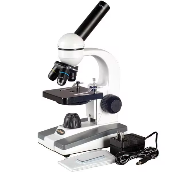
Ethical and Environmental Considerations in Compound Microscope Use
As the demand for compound microscopes grows, so does the need to address ethical and environmental concerns. Researchers and educators must consider the impact of their practices on both the environment and the communities they serve.
Sustainable Practices:
- Energy Efficiency: Modern compound microscopes often feature LED lighting, which consumes less power than traditional halogen bulbs.
- Recycling Programs: Some manufacturers offer take-back programs to recycle old microscopes responsibly.
- Minimizing Waste: Using reusable slides and coverslips instead of disposable ones reduces plastic waste.
Ethical Implications:
- Access Equity: High-quality compound microscopes can be expensive, creating disparities in educational institutions. Initiatives like open-source microscope designs aim to democratize access.
- Animal Welfare: When using biological specimens, ensure ethical sourcing and compliance with regulations to avoid harm to ecosystems.
Community Engagement:
- Public Workshops: Hosting community events where people can explore compound microscopes fosters scientific literacy and engagement.
- Collaborative Research: Partnering with local schools or NGOs to share resources ensures broader impact and sustainability.
By adopting these practices, users of compound microscopes can contribute to a more sustainable and equitable scientific community.